- 48 Hour Chick Embryo Serial Cross Section Chart
- 48 Hour Chick Embryo Serial Cross Sectional
- 48 Hour Chick Embryo Serial Cross Section Diagram
- 48 Hour Chick Embryo Serial Cross Section
Chicken: 22-28 h. In the stage from 22 hours on, the somites formed in the mesoderm at the left and right side of the neural walls become visible. After 24 hours 4 to 5 segmented paired blocks can be discerned. Later on, these structures will differentiate into the vertebrae, the ribs, a part of the skin and the dorsal muscles. 48 Hour Chick Embryo Cross Section Rating: 4,3/5 5965 reviews. 48-hour Chick Embryo Small Image Menu Instructions: The images on this menu will take you to a series of sub-menus containing more small-sized images such as these.
 On this page the following view of the embryology in chicken are displayed:
Stage 48 hrs
| Whole mount preparation 48 hrs | |
|---|---|
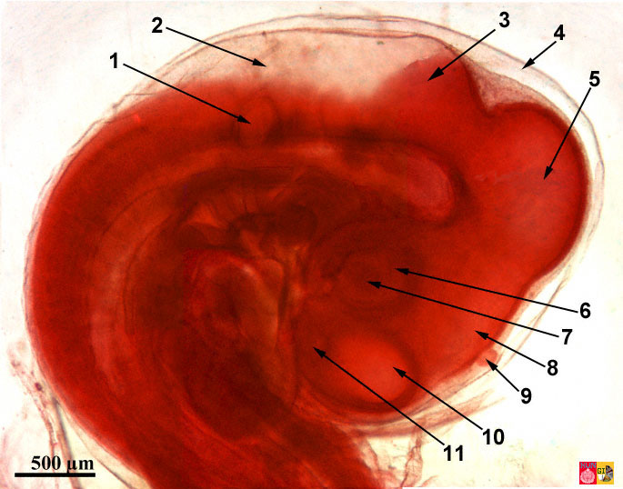
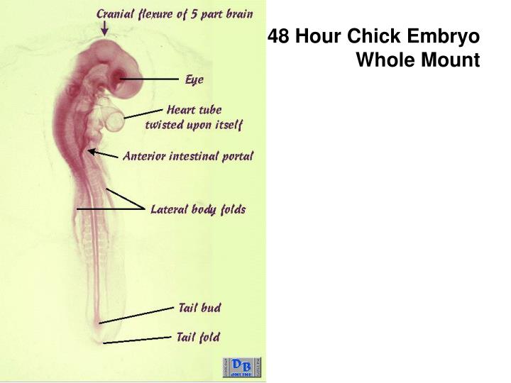
| Information: The position of the embryo with respect to the yolk changes strongly about 48 hours after fertilization. In addition to the head fold of the amnion, also the lateral and caudal amniotic folds begin to form. The outgrowth of the cranial flexure is so strong that the forebrain and hindbrain vesicles become almost located to each other. The cephalic region of the embryo is twisted in such a manner that the left side comes to lie next to the yolk. A second flexure appears at the transition of the head and the body just behind the heart region. The embryo takes now the shape of a C. The head becomes covered by a double fold. These folds definitely establish the first extra embryonic membrane (=outside of the embryo): the amnion membrane. The vitelline (yolk rich) arteries and veins become connected with the extra embryonic circulatory vessels. A few branchial grooves are already visible. The brain divides in to 5 vesicles: telencephalon and diencephalon (both formed by the division of the forebrain vesicle), mesencephalon, metencephalon and myencephalon (both formed by the division of the hindbrain vesicle). The lens placode (placode=plate) will form the lens vesicle, the optic vesicle will become the optic cup and the auditory placode the auditory pit. The heart differentiates in to 4 compartiments: the sinus venosus, connected with the veins, the atrium, the U-shaped ventricle and the bulbus cordis. The atrium and ventricle are well distinguishable in the figure. | |
| Embryology in chicken 48 hours after fertilization: stained whole-mount preparation 1 Amnion, 2 Metencephalon, 3 Mesencephalon, 4 Optic cup + lens, 5 Prosencephalon, 6 Otic vesicle, 7 Branchial arches, 8 Atrium , 9 Ventricle, 10 Lateral fold, 11 Lateral mesoderm, 12 Vitelline arteria / vein, 13 Somite, 14 Spine, 15 Tail fold | |

48 Hour Chick Embryo Serial Cross Section Chart
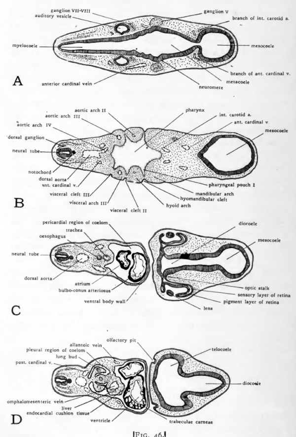
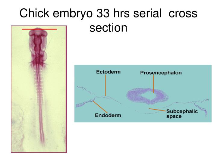
| Cross section chicken embryo - 48 hours | |
|---|---|
| A. Ear and eye formation: left whole mount and right cross section (dorsal part oriented to the left here) | |
| 1 = Myencephalon, 2 = Otic vesicle, 3 = Chorda, 4 = Pharyngeal cavity, 5 = Pharynx, 6 = Optic vesicle, 7 = Lens placode (lens formation), 8 = Optic cup, 9 = Diencephalon | |
| a, b, c, d : schematical representation of the eye formation overtime; e and f: detail of sections 1 = Neurectoderm, 2 = Epidermal (surface) ectoderm, 3 = Mesoderm, 4 = Optical vesicle, 5 = Optical cup, 6 = lens, lp = Lens placode (lens formation) | |
| B. Heart formation: left whole-mount preparation and right cross section (dorsal part oriented to the left) | |
| 1 = Spine, 2 = Somite differentiation (sclerotome dermamyotome), 3 = Chorda, 4 = Dorsal aorta arches, 5 = Cranial cardinale vein, 6 = Pharynx, 7 = Pericardial cavity, 8 = Heart formation | |
| C. Heart formation: left wholemount preparation and right cross section (dorsal part oriented left) | |
| 1 = Spine, 2 = Dermatome, 3 = Chorda, 4 = Dorsal aorta, 5 = Foregut, 6 = Truncus arteriosus, 7 = Atrium, 8 = Ventricle, 9 = Heart region |

48 Hour Chick Embryo Serial Cross Sectional
Stage 55 hrs
48 Hour Chick Embryo Serial Cross Section Diagram

| Dextro dorsal view at 55 hrs according to Patten |
|---|
| Patten, B.M. (1920). The Early Embryology of the Chick. Philadelphia: P. Blakiston's Son and Co. |